Article Text

Abstract
Background Many studies have discussed acute compartment syndrome in children associated with or without fractures and have given their visible perspectives. Little is known about the nerve involvement and the factors associated with recovery patterns in these patients. We intend to propose that ischemic nerve and muscles tend to regenerate after surgical decompression but in a different pattern and the given circumstances.
Methods Twenty-four children with acute compartment syndrome in the upper limb were analyzed between 2009 and 2015. Data included demographic features of these patients, the time interval between the injury and surgery, and the attempt to correlate with motor and sensory recovery.
Results The average follow-up was 67.3 months (range 59–80). Of the 24, 14 patients (58%) had immediate recovery of motor and sensory functions. The remaining 10 patients had variable recovery patterns with a mean time for the radial nerve, median nerve and ulnar nerve motor recovery of 6.0, 7.5 and 8.5 months, respectively, and sensory recovery at 12, 12 and 13 months, respectively. The overall study had a mean sensory recovery as per the Medical Research Council (MRC) of S3 in 3 (12%) and S4 in 21 (88%). The mean 2-point discrimination (2PD) was 6.9 mm (range 5–10). Twenty-one patients (88%) had a full range of movements at their final follow-up with a mean Visual Analog Scale score of 0.6; a quick Disabilities of the Arm, Shoulder and Hand score of 5.9 (range 2.3–25.0) and a Mayo wrist score of 79.
Conclusions There was a definite motor and sensory recovery in patients who underwent surgical decompression in acute compartment syndrome of the upper limb irrespective of age, gender, delay in presentation and various etiologies. The motor and nerve fibers can regenerate after ischemic sequela of compartment syndrome.
- orthopedics
This is an open access article distributed in accordance with the Creative Commons Attribution Non Commercial (CC BY-NC 4.0) license, which permits others to distribute, remix, adapt, build upon this work non-commercially, and license their derivative works on different terms, provided the original work is properly cited, appropriate credit is given, any changes made indicated, and the use is non-commercial. See: http://creativecommons.org/licenses/by-nc/4.0/.
Statistics from Altmetric.com
Key messages
What is already known about this subject?
Acute compartment syndrome in the forearm of children is an orthopedic emergency that needs urgent care to prevent irreversible nerve damage, muscle fibrosis, atrophy, contractures and deformities.
The presentation of children with acute compartment syndrome is unique in many ways, including undue delay, unreliable presentations of pain, paresthesia, pallor, pulselessness and paralysis and capacity to communicate because of anxiety, agitation and increased analgesic requirements.
Early decompression of the affected forearm is mandatory to prevent paralysis and contractures.
What are the new findings?
We found the statistical significance of tight oil bandage association with paralysis and high-energy mechanism of RTA/crush or fall presenting with severe pain and paralysis.
Single-incision fasciotomy was done to decompress all compartments, and the median and ulnar nerves were severely compromised at their entrapment sites in the wrist, forearm and elbow. Hence, adequate neurolysis from the carpal tunnel to the cubital tunnel should be done in all patients.
There was a statistical significance (p<0.5) assessed by Fischer’s exact test for the association between intervention <24 hours and >24 hours after injury of compartment syndrome to functional outcomes.
The mean times for radial, median nerve, and ulnar nerve motor recovery were 6.0, 7.5 and 8.5 months, respectively. The sensory recovery was in the proximodistal direction. It was complete recovery in the forearm at a mean of 10 months after injury (range 7–12) and in the hand at 13 months (range 10–14).
Key messages
How might it impact on clinical practice in the foreseeable future?
Our study proves that surgical decompression plays a vital role in the recirculation of blood supply to the ischemic nerves and muscles, thereby preventing muscle fibrosis, atrophy, contractures and deformities. We also proved that fibrotic muscle regains recovery over some time (6–12 months) based on radial, median and ulnar motor recoveries, producing full and excellent functional recovery in 21/24 (88%) children. The sensory recovery (proximodistal) was also seen in all patients (100%) with an average 2-point discrimination of 6.9 mm and an Medical Research Council grade of S4 in 88% and S3 in 12%.
Introduction
Acute compartment syndrome in the forearm of children is an orthopedic emergency that needs urgent care to prevent irreversible nerve damage, muscle fibrosis, atrophy, contractures or deformities.1 2 The presentation of children with acute compartment syndrome is unique in many ways, including undue delay, unreliable presentations of pain, paresthesia, pallor, pulselessness and paralysis (5 Ps) and capacity to communicate, owing to anxiety, agitation and increase in analgesic requirements.3 4 Early decompression of the affected forearm is mandatory to prevent paralysis and contractures.5 Volkmann’s contracture is defined as the end result of an ischemic injury to the muscles and nerves of the limb. Volkmann’s ischemia is the acute episode of pain, aggravated by passive stretching and neurological deficit resulting from ischemia of muscle and nerve.6 The extent of muscle recovery varies with the completeness of the destruction of muscle tissue and is directly related to the extent of motor recovery. Muscle regeneration seems to occur from a small number of subfascial fibers that survive the ischemic episode; regeneration is seen to be active even later than 76 weeks after injury.7 Several aspects of nerve and muscle recovery following fasciotomy have not been investigated. There seem to be associations between various additional factors affecting the functional outcomes in these patients. We propose that ischemic nerve and muscles tend to regenerate after surgical decompression but in a different pattern. Also, we analyze different factors contributing to functional recovery indirectly reflecting the ischemic nerve and muscle recovery.
Methods
Patients and statistical analysis
We conducted a retrospective analysis of 24 children admitted between 2009 and 2015 with different features of acute compartment syndrome in the upper limb. Patients with a head injury and lower limb injuries were excluded from this study. All patients had basic blood investigation and radiographs of the involved upper limb. In addition, tests for myoglobinuria, troponin and blood lactate were done to investigate for late/missed compartment syndrome in these patients. Based on the arrival of the emergency with clinical features and the surgeon’s discretion, 8 patients had compartment pressure measured, and the remaining patients were wheeled into the operating room at the earliest without compartment pressure management. Each patient’s data for the study were collected at the hospital, and pictures were taken with the appropriate consent. No patient was lost to follow-up. Based on the age, injury pattern, associated soft-tissue injuries/fractures, the time interval between the injury and admission and the time interval between the injury and surgery, motor and sensor recovery of these children with their functional outcomes were interpreted. Qualitative (gender and side) and quantitative variables (age) were evaluated using t-test. A comparison of the variables was done using χ2. Fischer’s exact test was used to compare the functional outcome in patients with a time of <24 hours from injury to fasciotomy to patients with a time of >24 hours to fasciotomy. Two-tailed p values of 0.05 or less were considered significant.
Surgical technique
All patients had single-incision fasciotomy as described by Henry (figure 1) under general anesthesia. The incision was extended proximally with slight curvilinear across the elbow flexion crease (oblique) and distally at volar wrist crease (Bruner) to decompress the lacertus fibrosus/antecubital fossa contents and the carpal canal, respectively. The advantage of the ulnar-side approach was that flexor tendons and ulnar and median nerves were left with good soft-tissue coverage in the area where necrosis was most likely to occur. Depending on the intraoperative findings and surgeon discretion, primary wound closure and delayed primary closure with or without skin grafting were done.

Schematic diagram showing the skin incision for surgical decompression for releasing carpal tunnel in the distal and up to cubital tunnel proximally if needed (drawn by the author).
Case 1
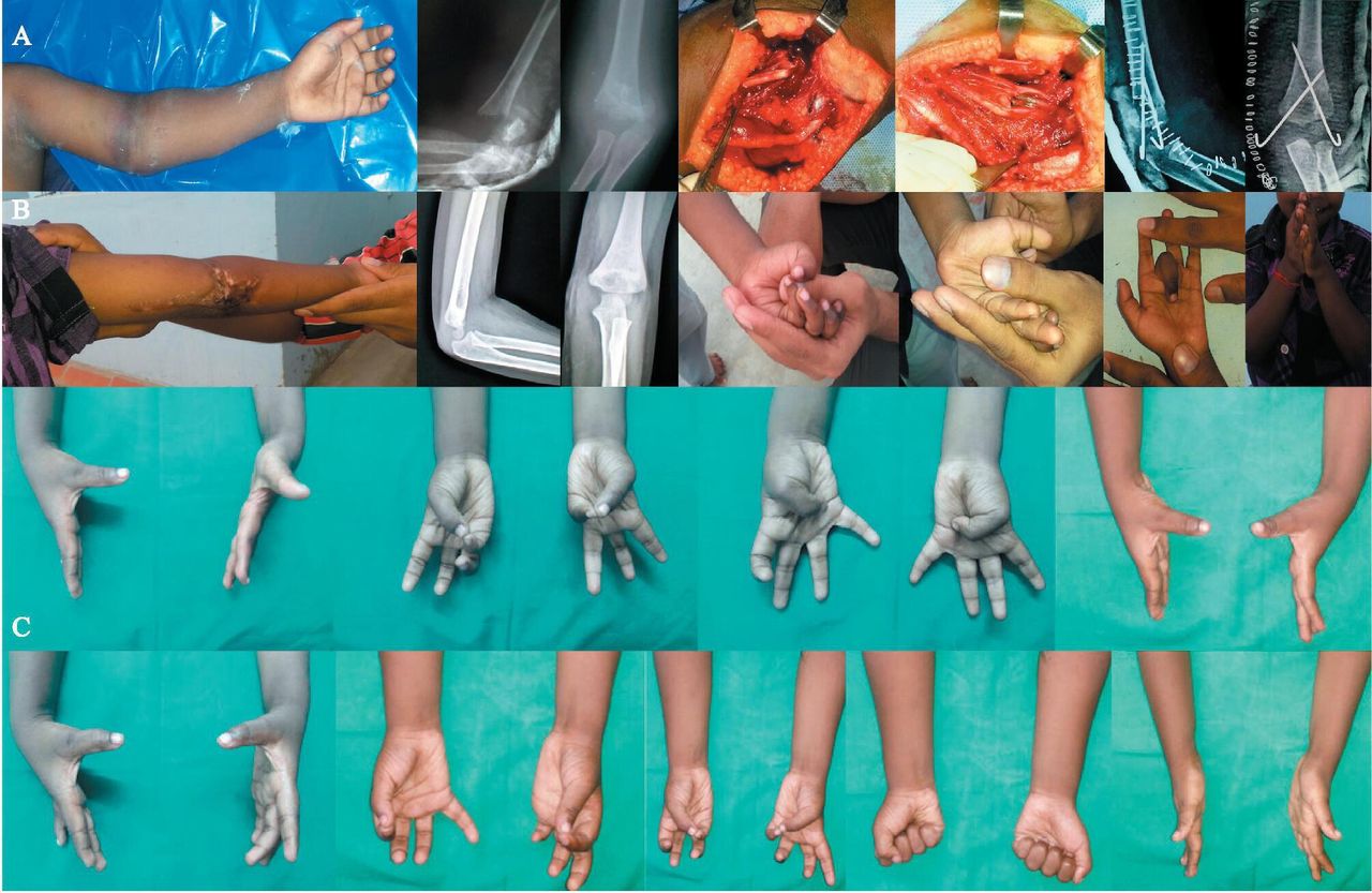
A boy in his early childhood who had a road traffic injury and accidentally fell, injuring his left upper limb, went to a non-physician to have a tight oil bandage on the same day. He came to us 6 days after the injury with numbness and paralysis of the entire upper limb. Radiographs of the left elbow showed an intra-articular olecranon fracture. Examination revealed complete motor and sensory loss in the entire upper limb with warm hand and good capillary refill. Considering the clinical findings and tense forearm, he was taken for immediate fasciotomy through the volar incision (Henry’s). Adequate decompression was done. Delayed primary closure was done at the fifth postoperative day (figure 2A). Minimal wound gaping was seen during the follow-up, and the wound healed with regular dressings. Wrist and finger extension was seen in the immediate postoperative period with a claw hand. He was put on regular passive therapy and claw splints in between therapy. At 8 weeks, the claw deformity improved with median (flexor digitorum superficialis, FDS) and ulnar nerve innervated muscles (finger abduction/adduction) recovering (figure 2B), and the skin had multiple raw areas around the suture lines. Median and ulnar nerve recovery was complete at 8 months of follow-up. He had excellent functional recovery with a Visual Analog Scale (VAS) score of 0, a quick Disabilities of the Arm, Shoulder and Hand (DASH) score 2.3 and a Mayo wrist sore of 85, and returned to school at 9 weeks. Sensory recovery was S4 with 2-point discrimination (2PD) of 5 mm at the final follow-up (figure 2C,D).

Intraoperative, clinical and follow-up pictures of an 8-year-old boy. (A) Intraoperative pictures showing adequate surgical incision and decompression of both median and ulnar nerves in an 8-year-old boy who came to the emergency room 6 days after the initial injury with complete paralysis of the upper limb. (B) Clinical pictures of motor recovery and multiple raw areas around the suture lines. Recovering median and ulnar innervated muscles showing a combined claw hand picture. (C) Full functional motor recovery at 8 months after surgery with wounds healed. (D) Full functional motor recovery (median and ulnar nerves) at 4 years following fasciotomy.
Case 2
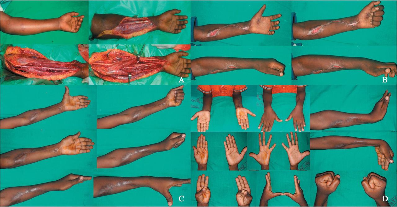
A boy in his middle childhood fell from his father’s bike and injured his left elbow. He was taken immediately to a hospital, where radiographs were not taken, and instead he was given analgesics. Despite a high dose of oral analgesics, he had severe pain, swelling and subsequent weakness and paralysis. All of this occurred about 1 day after injury, and the boy presented in our emergency room with complete motor and sensory loss. The limb was pink but pulseless. Serum myoglobinuria, lactate level and troponin were found within normal limits. Radiographs showed a displaced supracondylar (SC) humerus, which was stabilized and fixed with Kirshner wires (K wires). The thrombosed small segment brachial artery was excised, and an end-to-end microsurgical anastomosis was done. Fasciotomy was done, and the wound was closed primarily (figure 3A). He subsequently developed superficial skin necrosis, which settled with regular dressings. He developed claw hand (combined median and ulnar nerve involvement) in the immediate postoperative period (figure 3B) and was put on aggressive hand therapy with splints in between. He regained motor and sensory recovery with a full range of movements at the seventh month of follow-up. His sensation was normal S4 with 2PD 6 mm at the final follow-up (figure 3C).
Preoperative, clinical, intraoperative, nerve recovery pattern of a 5 year-old boy following a tight oil bandage treatment. (A) A 5-year-old boy who had fell from height managed initially with tight oil bandages came to us >24 hours after injury. Radiographs revealed displaced SC fracture humerus. The upper limb with pink, pulselessness and paralyzed. SC fracture was reduced and stabilized with K wires. Brachial artery was repaired. Fasciotomy was deferred based on the intraoperative findings of the supple forearm. (B) Postoperative pictures showing recovering FDS (median nerve), finger flexion on MCP joint flexion (ulnar nerve) with radiographs showing healed SC fracture with unstable scar over the elbow. (C) Full functional motor recovery (median and ulnar nerve) at 3 years following fasciotomy. FDS, flexor digitorum superficialis; K wires, Kirshner wires; MCP, metacarpophalangeal; SC, supracondylar.
Results
The most common mode of injury was road traffic injury and fall over the hand. There were 19 male children with a mean age of 8.6 years and 5 female children with a mean age of 8 years (tables 1 and 2). Thirteen children had right-side involvement. The age (p=0.23), side involved (p=0.64) and gender (p=0.52) had no statistical significance to outcomes in this study.
Demographics and treatment
Functional recovery and outcome
Etiology
Most of the patients developed compartment syndrome secondary to high-energy mechanisms of injury, with the most common being road traffic accident (RTA) in 17/24 (70%) of the cases. Five children had fallen from height, injuring the upper limb and subsequently developing compartment syndrome in the setting of fracture. Two children had intravenous infiltrations during their stay in the intensive care unit (ICU) and developed compartment syndrome with pressure of >40 mmHg. The mode of injury did not have significance to the outcome in this study (p=0.73).
Clinical presentation
Classically, a compartment syndrome diagnosis is associated with the 5 Ps. Severe pain was the initial presentation in our study in 19/24 patients (76%) associated with one or more 5 Ps. However, all patients had paralysis of the upper limb (complete motor and sensory loss) at presentation in the emergency room. We found no statistical significance of tight oil bandage association with paralysis and high-energy mechanism of RTA/crush or fall presenting with severe pain and paralysis (p>0.05). Two patients who were getting treatment in the ICU were referred for sudden onset of forearm swelling around the intravenous infusion area. They were operated on within 6 hours of the incident and had an excellent functional outcome at the final follow-up (table 3).
Correlation of multiple variables with full/good functional recovery versus partial/poor functional recovery
Tight oil bandage and undue delay in presentations
A special focus of attention towards this native oil bandage, which was applied by the non-physicians in the near vicinity, was the reason for the delay in presentation to the hospital. An oil bandage was traditionally used to treat fractures by the local people that had no medical background and had limited access to medical care. They applied oil and tried to set the bone with tight bamboo sticks or native dressings, which could have triggered the vicious cycle of compartment syndrome leading to development of transient/irreversible ischemic damages to nerve and muscle (n=7). All of these children had a trivial injury and sustained undisplaced olecranon fractures (n=2), SC fractures (n=2), radial head (n=1), proximal ulna (n=1) and distal radius fracture (n=1). The average time between injury and admission was 82 days (range 1–144) in these patients. Single-incision fasciotomy was done to decompress all compartments. Intraoperatively, the median and ulnar nerves were severely compromised at their entrapment sites in the wrist, forearm and elbow. Hence, adequate neurolysis from carpal tunnel to the cubital tunnel was done in all these patients.
Brachial artery injury was associated with paraesthesia and pain, with or without other 5 Ps in our study. The presence of these two symptoms confirmed the onset of ischemia sequela and compartment syndrome, producing motor and sensory deficits. Six out of seven children (olecranon, n=2; SC humerus, n=1; with brachial artery, n=1; radial head, n=1; proximal ulna, n=1; distal radius, n=1) with a tight bandage and delay in presentation had immediate recovery of radial nerve, but claw deformity persisted.
Nerve involvement and recovery
Of the 24 patients, 14 patients (58%) had immediate recovery of motor and sensory functions. Ten (42%) patients had varied patterns of recovery of radial, median and ulnar nerves, of which, three (13%) had radial, median and ulnar nerve palsies, six (25%) had combined median and ulnar nerve palsies, one patient (4%) had median nerve injury alone.
1. Radial nerve
The mean time to appreciate thumb extension (extensor pollicis longus) was 12 weeks (range 8–16), and the sensation was 24 weeks (range 16–32). The motor recovery was complete at a mean of 6 months (range 5–8) after injury. The sensory recovery was complete in the forearm at a mean of 7.5 months (range 6–9) after injury and in the hand at 12 months (range 10–13).
2. Median nerve
The mean time to appreciate FDS flexion in fingers was 10.3 weeks (range 2–20) and the sensation was 13 weeks (range 2–20). By 3 months, all of them had claw hand, which explains the common involvement with the ulnar nerve. The motor recovery was complete at a mean of 7.5 months (range 6–9) after injury. The sensory recovery was complete in the forearm at a mean of 10 months (range 7–12) after injury and in the hand at 13 months (range 10–14).
3. Ulnar nerve
The mean time to appreciate finger abduction/adduction was 12 weeks (range 2–26), and sensation was 19 weeks (range 2–26). The motor recovery was complete at a mean of 8.5 months (range 7–12) after injury. The sensory recovery was complete in the forearm at a mean of 10 months (range 7–12) after injury and in the hand at 13 months (range 10–14).
Among the chidrens with acute compartment syndrome and nerve injuries, one patient (4%) who had been treated elsewhere for displaced SC humerus fractures had superficial and deep infection. No motor or sensory recovery was seen immediate to the surgery. Partial radial nerve (active thumb extension) recovery was seen at 12 weeks, median nerve at 16 weeks and ulnar nerve at 20 weeks. Despite this recovery, the patient still had claw hand and stiff metacarpophalangeal and proximal interphalangeal joints.
One patient (4%), who came 171 hours after injury and had K wires driven through ulnar nerve and radial nerves, developed septic arthritis, triggering compartment syndrome. The abscess was drained, and the K wires were removed with adequate decompression and neurolysis. Postsurgery, there was no immediate motor and sensory recovery. Partial radial nerve recovery was seen at 16 weeks, median nerve recovery at 20 weeks and ulnar nerve at 26 weeks with a stiff elbow and stiff claw hands.
The average sensory recovery as per Medical Research Council (MRC) was S3 in 3 (12%) and S4 in 21 (88%). The average 2PD in our entire study was 6.9 mm (range 5–10).
Compartment pressure measurement
Based on the clinical findings and surgeon’s discretion, compartment pressure was not measured in 16 children. These children presented to the emergency room with acute compartment syndrome with motor and sensory paralysis (n=13), brachial artery injury with paralysis (n=2) and intravenous infiltrate in one child in the ICU. All of them were wheeled into the operating room at the earliest. had a compartment measurement recorder and were found to have >30 mmHg. There was no statistical significance (p=1.0) for the association between clinical assessment and compartment pressure measurement test to functional outcomes (table 3).
Time to intervention
The average time interval between injury and admission in this study was 44 hours (range 5–192), and the average time interval between admission and surgery was 54 minutes (range 45–65). Eleven patients underwent surgical decompression >12 hours after injury, and 8 patients (73%) had excellent motor and sensory nerve recovery. Among these patients, seven had a delay because of native tight oil bandage. There was a statistical significance (p<0.5) assessed by Fischer’s exact test for the association between intervention <24 hours and >24 hours after injury of compartment syndrome to functional outcomes. Fifteen patients (63%) who were operated on within 24 hours after injury had excellent functional outcomes. Of the 9 patients who were operated on >24 hours after the injury, 6 (67%) had excellent motor and sensory recovery and 3 (33%) had good sensory recovery and partial motor recovery.
Fasciotomy and wound closure
Single-incision fasciotomies were performed in all patients as a definitive treatment of compartment syndrome. Two patients who had very late presentation (more than 7 days after initial closed reduction and K wire for SC humerus fracture with motor and sensory deficits) had wider surgical incision extended to the arm. Overall, 14 patients had primary wound closure with skin suturing. Five patients required delayed primary closure at an average of 5 days. Five patients also needed skin grafting at an average of 5 days, considering the wound status. None had skin grating failure in our study. Of the 14, who had primary wound closure, two patients had superficial skin necrosis with multiple small areas in the follow-up that healed subsequently without any interventions. The average stay in the hospital was 7.3 days (range 3–19). Patients undergoing primary closure had a hospital stay of an average of 6 days when compared with delayed closure with or without skin grafting of 11 days. One patient (4%) required debridement and drainage of the abscess and multiple surgeries for his left elbow operated on elsewhere with features of septic arthritis and stayed at the hospital for 19 days receiving intravenous antibiotics and dressings.
Functional outcome
The mean follow-up was 67.3 months (range 59–80). Twenty-one patients (88%) had a full range of movement at their final follow-up with a mean VAS score of 0.6; quick DASH score of 5.9 (range 2.3–25.0); and Mayo wrist score of 79. Full functional recovery, defined as the ability to return to preinjury levels of activity, was achieved in 21 of 24 patients (88%) in a mean time of 14 weeks (range 9–24).
Associated injuries
There was a statistical significance to the outcome in the children with associated injuries involving SC humerus fractures in our study (p<0.05). This was because of its association with K wire-related nerve injuries and infection sequela leading to partial motor nerve recovery with good sensation (S3).
Discussion
There have been few publications about the diagnosis and treatment of acute compartment syndrome in children.8 9 The most common site involved is pedestrian injuries to the distal tibia followed by high-energy injuries to the upper limb.10 Various prognostic factors, such as age, gender, injury pattern, location and the clinical presentation, are discussed in the literature.11 12 In our study, age (p=0.11), gender (p=0.52), side (p=0.64), mode of injury (p=0.73), etiology (p=0.86) had no significant impact on the outcome. Supracondylar humerus, time delay >24 hours, clinical presentation had significant impact on the outcome (p<0.05) (tables 3 and 4).
Comparative study and literature review about acute compartment syndrome in the upper limb
There has been no mention of the nerve involved in the upper limb compartment syndrome and its recovery pattern. This became the prime focus of our study, in addition to other vital information. Intracompartmental pressure (ICP) measurement is recommended in young children, unconscious patients, patients with regional nerve blocks and when the clinical signs are equivocal. It may not be necessary if the diagnosis is clinically evident.13
The traditional hallmarks of vascular insufficiency include 5 Ps; however, acute compartment syndrome in children may not have all of these classic features, making the 5 Ps less reliable.4 The diagnosis of compartment syndrome, therefore, should not be excluded based on the false assurances gained from the absence of these clinical findings.12 In our study, all of the patients had complete motor and sensory paralysis at the time of admission. Totally, 76% of our patients had pain as one of the initial complaints, in addition to a few of the other 5 Ps. Delay because of tight oil bandage by the traditional bone setter had paralysis (29 %) as the prime complaint, which is statistically significant in our study (p<0.05). Further, the presence of pallor, paraesthesia, paralysis and pulselessness indicates prolonged ischemia and more advanced disease.2 3
Our studies (table 3) have found that age, gender, fractures or soft-tissue injuries (intravenous infiltrate) have no statistical significance (p>0.05) in terms of good or poor functional recovery. Prasarn et al found favorable outcomes in 6/14 patients operated on within 6 hours of injury.9 We found statistically significant (p<0.05) and excellent functional improvements in 15 patients (63%) operated on within 24 hours after injury. Three (13%) patients operated more than 24 hours after injury had good sensory recovery and partial motor recovery. SC humerus fractures associated with late presentation, oil bandage and iatrogenic K wires injuring nerves had statistically significant poor functional outcome.
What’s more interesting in our study is the re-emergence of tight oil bandage in children with a paradigm shift in functional recovery. Sundararaj and Mani found tight bandage by the traditional bone setter to the upper limb produces irreversible motor damage, causing various degrees of contractures, (mild, moderate and severe), wasting of forearm muscles with claw, and ape-thumb deformities.14 None had any surgical intervention. None of his patients had a motor recovery. He absorbingly noticed that sensory recovery in the forearm happened at 6 months and in the hand at an average of 12 months and had no reason except nerve regeneration (table 4).15 16
In our study, tight oil bandage (29%) to the upper limb was associated with various fractures and caused motor and sensory paralysis. The average time between injury and admission was 82 days (range 1–144) in these 7/24 patients. Despite the delay, all seven patients underwent fasciotomy to have full functional recovery in (6/7) 86% of patients. Fifteen patients (63%) who were operated on <24 hours after injury and 67% of patients (6/9) who were operated on >24 hours after the injury had excellent functional outcome. The mean times for radial, median nerve and ulnar nerve motor recovery was 6.0, 7.5 and 8.5 months, respectively. The sensory recovery was in the proximodistal direction. It was complete recovery in the forearm at a mean of 10 months (range 7–12) after injury and in the hand at 13 months (range 10–14).
Our study proves that surgical decompression plays a vital role in the recirculation of blood supply to the ischemic nerves and muscles, thereby preventing muscle fibrosis, atrophy, contractures and deformities. We also proves that fibrotic muscle regains recovery over time (6–12 months) based on radial, median and ulnar motor recovery producing full and excellent functional recovery in 21/24 (88%) children. The sensory recovery (proximodistal) was also seen in all patients (100%) with an average 2PD of 6.9 mm and with an MRC grade of S4 in 87% and S3 in 13% (range 10–14 months after surgery) (tables 1 and 2). Totally, 12% of children had partial motor recovery despite multiple surgeries for soft-tissue infection, osteomyelitis and stiff joints in the subsequent days, compromising the nerve and muscle recovery but recovering S3 sensation. Both motor and sensory recoveries are from the newly regenerated nerve supply. Thus, our hypothesis was confirmed by the fact that motor fibers and sensory fibers after fasciotomy regenerated to produce 100% sensory recovery and 88% motor recovery in our patients.
The main limitations of our study were the small sample size and retrospective design. Since the nerve recovery does not follow Wallerian degeneration, axonal regeneration or end-organ reinnervation patterns, a histopathological examination can throw light on the recovery details. The muscles can be taken up for histopathological evaluation while doing fasciotomy to look for its vascularity, viability and regeneration. Again, the muscle may have a different level of ischemic injuries either completely or partly ischemic, so this becomes quite uncertain. Injury to the nerves can coexist with brachial artery injury or with compartment syndrome, and the clinical entities may overlap, making the diagnosis frequently difficult if not impossible. The onset of motor and sensory paralysis as in our cases sequel to tight oil bandage; the recovery pattern of median and ulnar nerve at a mean of 8.0 and 8.5 months rules out neuropraxia/axonotmesis type of injury. Again, for a rational explanation, the different pattern of neuromuscular recovery needs a large prospective study containing in vitro models, in vivo nerve injury models, molecular expressions and neuromuscular junction studies. It is logical to understand that ischemia induces markedly elevated ICP, which gets decompressed by fasciotomy and allows the nerve and muscle to regenerate back in their due course of time.
Conclusions
Surgical decompression in acute compartment syndrome involving the upper limb in children allows the recirculation of blood and muscle and nerve fibers to regenerate definitely to produce excellent proximodistal sensory recovery and full functional motor recovery irrespective of various contemporary factors. Early surgical decompression produces excellent functional results.
Acknowledgments
The author acknowledges the significant contribution of Dr Selvam, who is an associate professor in Social and Preventive Medicine, Government Medical College, for his statistical analysis in this study.
References
Footnotes
Twitter @terrencejose
Contributors JTJ was solely responsible for the design, data collection and analysis, and writing of the manuscript.
Funding The authors have not declared a specific grant for this research from any funding agency in the public, commercial or not-for-profit sectors.
Competing interests None declared.
Patient consent for publication Parental/guardian consent obtained.
Ethics approval The study observed medical ethics standards and was approved by ethical committee board of Olympia Hospital & Research Centre (no. 02/2020).
Provenance and peer review Not commissioned; externally peer reviewed.
Data availability statement Data sharing not applicable as no datasets were generated and/or analyzed for this study. Data can be available but not the patient’s pictures and details as these require the patients' consent and their rights to accept/deny.